This guide breaks down the purpose of eye exams, how frequently you should conduct eye exams, the necessary components of eye exams, and the method of preparing for eye exams. Know the average duration for an exam, the cost, and the recent technological advancement of eye exams. Get the various types of specialized eye exams and the conditions detected during eye exams.

This is several tests done by an optometrist or ophthalmologist to measure your vision acuity and also to detect eye-related conditions and diseases that might be affecting the overall health of your eyes. It is a comprehensive evaluation of eye health. An eye exam is also known as a routine eye exam. It is the regular check to know the overall status of your eye health.
It involves various tests to detect eye conditions. These can be eye defect conditions, such as cataracts, myopia, hyperopia, astigmatism, etc. This exam can check unrelated medical problems such as diabetes that cause symptoms that affect the structures of the eyes.
An eye exam is done by an optometrist or ophthalmologist in case there are any specific things about your eyes that need to be addressed.
Eye exams should be done regularly, even if you don’t have eye problems that need treatment or monitoring. It helps you know your eye status and maintain overall health.
This guide breaks down
Purpose of Eye Exam
The purpose of eye exams is for your eye doctor to assess the overall health status of your eyes. The optometrist checks the following:
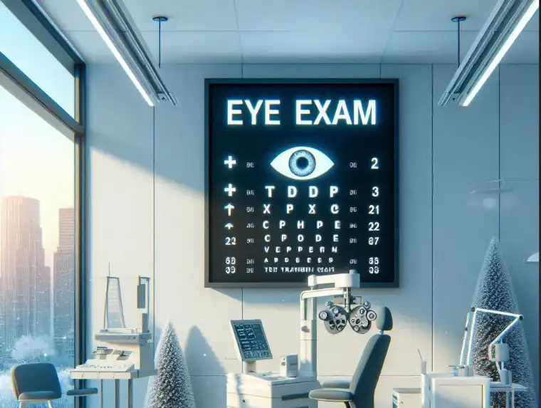
- Visual acuity: This assesses how clearly your eye can see. And it is mostly done using Snellen Chart. In the chart, large symbols or letters are at the top, while it progressively get smaller down the chart.
- Eye muscle movement and alignment: This checks if your eyes align well and move correctly in all directions without moving your head.
- Color vision
- Visual fields: This assesses how much you see on each side of your view without moving your eyes. It detects gaps or blind spots that shouldn’t be there.
- Refractive error: It detects how light waves pass through the cornea and lens of the eye.
- The health of the cornea
- Risk of glaucoma
- Surrounding outer tissue (ocular adnexa): The ocular adnexa includes all the parts of your eye and face that aren’t the eyeball itself, e.g., lashes, eyelids , tear system, lymph nodes, etc. In this section, your eye doctor assesses the specific eye parts for signs that they’re working.
Frequency of Eye Examination
The frequency of eye examinations depends on many factors. These factors may include the individual’s age, past eye history, family history of eye disease, medical history, and symptoms or ocular findings encountered. If there was an eye-related disease detected, the frequency of eye examination will depend on how severe the conditions are, the response to treatment (or surgery), and the potential for detecting progression of the abnormality.
According to the Canadian Association of Optometrists (CAO), they accepted the recommendations contained in the “Review of the Canadian Association of Optometrists Frequency of Eye Examinations Guidelines: An Evidence-Based Approach.”
The guidelines can help an individual determine the need for an eye examination. The guideline stated that the minimum frequency of examination for those at low risk is as follows:
Infants and Toddlers (birth to 24 months) Infants and toddlers should undergo their first eye examination between the ages of 6 and 9 months.
Pre-school Children (2 to 5 years) Pre-school children should undergo at least one eye examination between the ages of 2 and 5 years.
School-age children (6 to 19 years) should undergo an eye examination annually.
Adults (20 to 39 years) Adults aged 20 to 39 years should undergo an eye examination every 2 to 3 years.
Adults (40 to 64 years of age) should undergo an eye examination every 2 years. Adults (65 years of age or older) should undergo an eye examination annually.
For young individuals at higher risk for certain diseases, such as African-Americans who are at higher risk for glaucoma, comprehensive eye examinations should be done every 2 to 4 years for those under age 40, every 1 to 3 years for those aged 40 to 54, and every 1 to 2 years for those aged 55 to 64, even in the absence of visual or ocular symptoms.
Components of Eye Exam
Visual Acuity
A visual acuity test shows how well your eyes can see. That is the degree of your visual clarity. The Snellen chart is used for this test. This chart is made up of 11 rows of capital letters or symbols, each row written in a descending size. You read the letters from 20 feet away from where you are sitting or standing. The smallest letters you are able to read will determine your accuracy. If your vision is normal, your visual acuity may be written as 20/20. Vision of less than 20/20 means you have hyperopia or myopia.
Refraction testing
In refraction testing, it is a subjective test to measure nearsightedness, astigmatism, presbyopia, and farsightedness. Your eye doctor uses a computer-guided refractor to know how light rays focus at the back of an individual’s eye. Retinoscopy is another tool used in this diagnostic process. Your eye doctor will shine a light into the depths of the eye to assess the movement pattern of your eye, which is known as the “red reflex.” If the light reflects back by your retina, the eye doctor records the degree of the refractive error. Corrective lenses or even refractive surgery can be prescribed if there is a notable deviation from normal refraction.
The doctor places a phoropter, an instrument that has varieties of lenses showing different degrees of vision correction on it, in front of your face.
The refraction test is used by the eye doctor to develop your contact lenses or eyeglass.
Pupils Dilation
It is a common procedure in eye examination. The dilation and constriction of the pupils in response to light reveal the health of the eye and also diagnose different conditions. Pupillary reactions can reveal neurological problems.
The acronym PERRLA is used to describe the findings of a pupillary response test. It stands for Pupils Equal Round Reactive to Light and Accommodation—the ability of the eyes to focus on objects that are close-up and far away. After the end of an exam that results in blurred vision, allow some time for the blurred vision to wear off before going back to your normal activities. Always go with your sunglasses because recently dilated eyes are sensitive to light.
Slit Lamp Examination
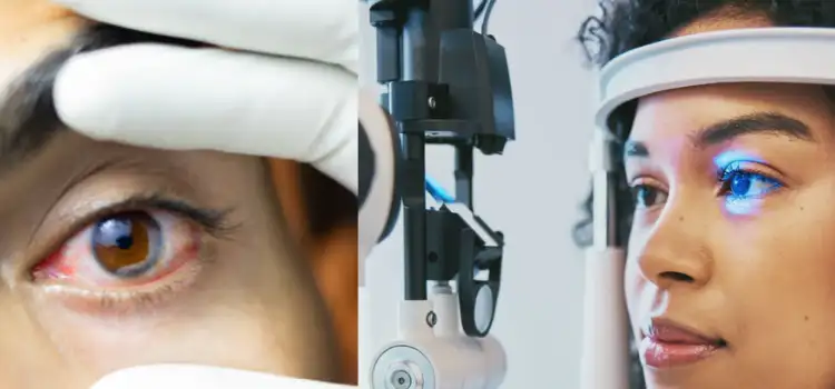
This examination is done to examine the front and back of your eye as part of an overall test of general health. The slit lamp or biomicroscope is the tool used by the doctor during this eye exam. It is a lighted microscope for examining the eye. It helps the eye doctor to easily see the eye’s inner fluid chamber, including the conjunctiva, lashes, iris, lens, and cornea. Also, it both magnifies the eye many times and illuminates it with a bright light so individual structures can be examined., including the lids and lashes, conjunctiva (the membrane that lines the eyelid and white of the eye), This reveals any eye defects or diseases, e.g., cataracts.
Checking for glaucoma
A tonometer is used for glaucoma screenings. A tonometer measures fluid pressure or intraocular pressure, which indicates the risk of having glaucoma. The ophthalmologist performs applanation tonometry, which assesses the level of force necessary to momentarily depress the cornea. This process uses fluorescein eye drops administered with anesthetic. The eye doctor may also use a pachometer, which determines sound waves to gauge the cornea’s thickness.
Visual field test
A visual field test checks how well an individual can see objects both on your central and peripheral vision without moving the eyes. Your eye doctor will ask you to cover one eye, look ahead without moving the head or eyes, and indicate when an object comes into view.
You may be asked to focus on a point at the center of a screen and tell your eye doctor when the object changes location or moves off screen.
This test measures the muscles that control eye movement. It is usually a simple test conducted by moving a pen or small object in different directions of gaze.
Color blindness test
The test is done using a series of cards with multicolored dots (Ishihara color plates) arranged in patterns. This test is done to check your ability to differentiate shades of different colors from each other. Your eye doctor proceeds to diagnose different types and severity of color blindness.
Retinal Examination
It is also known as funduscopy or ophthalmoscopy. This is the last step in a comprehensive eye examination. The ophthalmologist conducts a thorough examination of the posterior part of the eye, particularly the optic disk and retina, as well as the choroid, the blood vessel layer that supports and nourishes the retina.
It starts with pupil dilation to get an optimal view of the structures of the eye, including the retina, optic nerve, blood vessels (choroid), vitreous, and macula. The dilated fundus examination is important during eye examination because the test can detect several eye diseases.
Can I do My Eye Exams at Home?
Yes, you can follow this guide to conduct your eye exams at home.
But you must be guided by a profesional eye doctor.
Preparing for an Eye Exam
Preparing for your eye exam does not require much but needs some homework before seeing your eye doctor. However, there are lists of some information that will make the appointment more successful and efficient for both you and your doctor. Here are some lists of information to make:
Write Your Symptoms and Concerns
Having a note of your symptoms and concerns ahead of the appointment can make you remember easily. This is because various screening tests on the arrival of your appointment can make you forget. There may be issues that brought you in, like eye strain, double vision, red eyes, or blurry vision. Or it may be that you just came in to check the overall health of your eyes.
Go With Your Current Prescription
When taking new measurements, it will be helpful to compare your last eyeglass prescription. Also take along your contact lenses; bring them in an unopened blister package or in their case.
Family medical history
Knowing your family‘s health history is important when assessing your risk of different diseases, especially various eye defects. If possible, get the information about your parents’ and grandparents’ eye health. Perhaps you know if they had glaucoma, astigmatism, macular degeneration, diabetic retinopathy, cataracts, or any other eye issue. Having this information is important for your eye doctor to assess your risk of developing the same condition.
Wear Sunglasses
Dilation of the pupil during an eye exam can make you light sensitive for some hours after the appointment. Wearing your dark sunglasses after the appointment can make your eyes feel more comfortable. Some offices may provide disposable sunglasses, but if you have your own, bring them along.
Duration
A comprehensive eye exam depends on the type of exam and your eye health; the testing will take about 30 minutes to an hour. If the eye exam will involve dilating your pupils, it could take longer because the drops need time to be effective.
An eye exam for contact lenses typically lasts longer than an eye exam to get glasses.
Cost of Eye Exam
Research made by the CDC noted that most adults don’t go for regular eye care checkups. This is because either they are unaware of their importance or concerned about costs. Financial support from your eye insurance company can make your eye exam less expensive.
According to Warby Parker, “the average cost of an eye exam without insurance is about $100.” But this figure is liable to change, depending on who you ask.
VSP, a prominent vision insurance provider, lists the national average cost of an eye exam without insurance at around $194.
The FAIR Health has a list of various estimates for different components of a check-up. An examination of your eyes for health issues is about $135, and refractive tests that determine your prescription are estimated at around $54. That means the average total cost of a comprehensive eye exam would be about $189.
Most people choose to go to optical retail stores, where the price of an eye exam is more affordable than going for an independent optometrist.
In general, eye exams range in cost from around $50 to $250, and the popularity of retail stores and optical chains gives the approximate average cost of $100.
With the aid of insurance, your eye exam will be less expensive. The cost will be about $10–$40 on average for a comprehensive eye exam—and that’s if your insurance doesn’t cover it entirely.
There are different factors that determine the cost of an eye exam without the help of insurance.
These factors include the type of eye care provider you visit, the kind of eyewear you’d like, and your location, which have an impact on the price.
Advanced Equipment for Eye Exam
Optical Coherence Tomography (OCT)

Optical coherence tomography, or OCT, is an ultrasound for the eye that makes use of light instead of sound waves to obtain high-resolution images of the drainage angle, optic nerve, and retina.
This equipment helps to image the surface of various tissues of the eye and also the underneath the surface. OCT uses a scanning laser to capture the image and will display the information in 3D, color-coded, and cross-sectional scans using computer software. OCT is better equipment for early diagnosis and continued management of glaucoma, macular degeneration, retinal detachments, and diabetic retinopathy. With OCT, serious nerve or macular conditions in cases of unexplained vision abnormalities can be ruled out.
The OCT scan does not need eye drops or a flash of light when taking the image. The images can be saved into your electronic health record for immediate viewing with your eye doctor and be available for future appointments.
Digital Retinal Imaging
Digital retinal imaging helps your optometrist evaluate the health of the back of your eye, the retina. It is important to know the health of the retina, optic nerve, and other retinal structures. Digital retinal imaging snaps a high-resolution digital picture of your retina. This picture clearly shows the health of your eyes and is used as a baseline to spot any changes in your eyes in future eye examinations.
Importance of Regular Eye Exams
Regular eye examination is important for knowing the overall health of your eye. They are also a beneficial part of detecting eye issues early to protect your vision. Some eye issues are unnoticed in their early stages. A comprehensive eye exam by your eye doctor can detect early stages of these eye diseases. Some reasons why you should go for an eye exam:
- Early Detection of Eye Diseases: Eye diseases like glaucoma, diabetic retinopathy, astigmatism, cataracts, and macular degeneration can be detected in their early stages during an eye exam, often before their symptoms are noticed and cause irreversible damage to your vision.
- Identification of Underlying Health Conditions: Some systemic diseases, such as diabetes, hypertension, etc., can be noticed by your eye doctor through changes in the retina or in blood vessels. Your eyes can provide an idea about your overall health. During an eye exam, the eye doctor may detect signs of systemic diseases, such as diabetes or high blood pressure, based on changes in the blood vessels or the appearance of the retina.
- Updating Prescriptions: For optimal vision, your eyeglass or contact lens prescription may be adjusted, especially if you’ve noticed changes in your vision.
- Prevention of vision loss: Regular eye exams can prevent vision loss or slow its progression by detecting and treating eye conditions early.
- Monitoring changes in vision: During regular eye exams, changes in vision can be monitored and addressed, ensuring that you have the appropriate prescription for glasses or contact lenses.

Eye Exams: Purpose, Cost, Conditions (AI Generated image for the purpose of illustration. (modified by author)
Conditions Detected During Eye Exams
Some eye diseases can be detected during a comprehensive eye exam. They include:
- Glaucoma: It is a severe eye issue that can lead to vision loss and blindness. It is also known as “silent thief of sight” because it has few early warning signs.
- Diabetes: the swelling and leakage of tiny blood vessels lined up to your retina can be a sign of diabetic retinopathy, an eye disease caused by diabetics. Sometimes, this can be the first sign of the disease condition.
- Cataracts: They are natural and frustrating, causing clouding in your eye’s lens. New corrective lenses are used for early treatment.
- Age-Related Macular Degeneration (AMD) is a condition that affects central vision. It is primarily caused by aging and can happen very slowly (dry AMD) or with a rapid decline (wet AMD). AMD happens in 3 stages and cannot be reversed.
- Blepharitis is a skin condition that causes inflammation of the eyelids. Bacteria on your eyelids build up, which leads to eye irritation. This can affect your vision and block your tear glands, resulting in dry eye disease.
Other diseases an eye exam could detect include:
- Heart disease
- Dry Eye Disease
- High blood pressure
- Thyroid disease
- Vitamin A deficiency
Follow-up Care
Attending follow-up appointments helps ensure a successful recovery and optimal vision correction. During these visits, adjustments to the treatment plan can be made by your eye doctor depending on your progress. The adjustments include modifying medication dosages, recommending additional therapies, or providing reassurance and support.
Follow-up visits are important for post-operative care and play a crucial role in achieving the best possible results for eye surgery patients.
Follow-up visits are important for monitoring healing, addressing complications, and ensuring proper vision correction after eye surgery.
Post-surgery medication and care are managed to enhance healing and prevent infection during follow-up.
During follow-up, your eye doctor addresses your questions and concerns, which provides personalized care and satisfaction.
Long-term eye health and maintenance are discussed and planned for during follow-up visits to promote overall well-being.
You build a strong patient-doctor relationship through effective communication and personalized care during follow-up visits.
Types of Specialized Eye Exams
Contact lens fitting exam
This is an additional test that will be performed in addition to a comprehensive exam if you want to or wear contact lenses. The exam is used for young adults and adults. These tests include:
- Tear film evaluation to ensure your eyes are good for comfortable contact lens wear.
- Cornea measurement.
- Assessment for the correct fit and type of contact lens for your eyes.
- Calculation of the correct contact lens prescription.
Pediatric exams
These eye exams are for young children and infants or toddlers. They involve several techniques and tests than for school-aged children or adults. Amblyopia is an eye condition that affects developing children. Taking them to an eye doctor for a routine exam is important because they are too young to explain what they can’t see well.
Binocular vision exam
A binocular vision exam may be done on school-aged children as part of a comprehensive eye exam. Adults having visual problems related to binocular vision may benefit from a binocular vision exam. A binocular vision exam will assess:
- Vision history, including any visual issues.
- Vision at distance, near, and intermediate ranges.
- Eye alignment and teaming.
- Ability to follow a moving object.
- Ability to focus up close and read for extended periods.
- Depth perception.
- Hand-eye coordination and spatial awareness.
Low vision exam
This eye exam is specialized for those with visual impairment. Low-vision patients do not have the ability to perform daily activities such as reading, driving, etc., even with glasses or contact lens correction. Low vision can be caused by decreased vision, decreased visual field, or both. It leads to a functional loss.
Diabetic Eye Exam
The eye exam is used to detect complications of diabetes, which is diabetic retinopathy. The risk of diabetic retinopathy increases the longer a person has diabetes. Uncontrolled blood sugar levels damage the blood vessels of the retina, causing them to swell and leak or even stop blood flow.

